皮内試験
いつ行うか?
- アトピー性皮膚炎の臨床診断が下され、関連アレルゲンを特定してアレルギーワクチンに組み込むことを希望する場合
- 犬におけるノミアレルギーを確定診断するための補助検査として
何を見つけるか?
- もっとも重要な環境アレルゲン(屋内棲息性ダニ(ハウスダストマイト);動物のふけ;樹木、雑草、芝の花粉;カビ)を特定する。

何が必要か?
- 通常約50種類の関連アレルゲン一式を、適切な皮膚検査濃度に希釈したもの
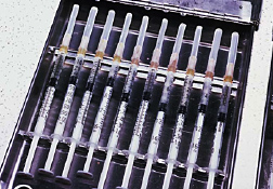
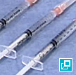
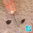
- ツベルクリンシリンジおよび25ゲージ針、バリカン、油性マーカーペン、反応部位を測定するためのノギスまたは定規
どのように行うか?
- 必要に応じて、患者をメデトミジンまたはプロポフォールで鎮静させる。
- 片側胸郭から被毛を刈り取る。
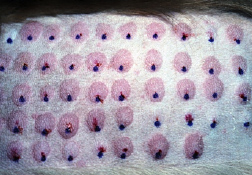
- 油性マーカーペンで注射部位に印をつける。
- 各アレルゲンと、陰性対照および陽性対照をそれぞれ約0.05 mLずつ注射する。
- 10~20分後に反応部位を確認して大きさ、紅斑、膨張の様子を評価し、0~4+のグレードを付ける。
ヒント
- 本試験は疾患を診断するものではなく、アトピー性皮膚炎の治療に免疫療法を用いる予定の患者にのみ行う。
- ノミアレルギーを有する犬の大部分は即時型反応を示すが、遅延型(24~48時間)反応しか示さないものもいる。
abscess
A discrete swelling containing purulent material, typically in the subcutis
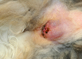
Perianal abscess in a dog
alopecia
Absence of hair from areas where it is normally present; may be due to folliculitis, abnormal follicle cycling, or self-trauma
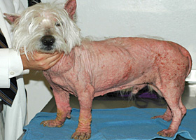
Extensive alopecia secondary to cutaneous epitheliotropic lymphoma
alopecia (“moth-eaten”)
well-circumscribed, circular, patchy to coalescing alopecia, often associated with folliculitis
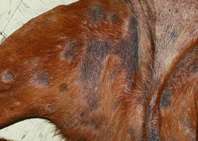
“Moth-eaten” alopecia secondary to superficial bacterial folliculitis
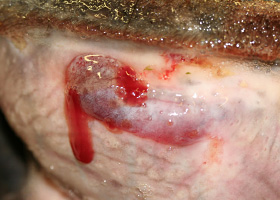
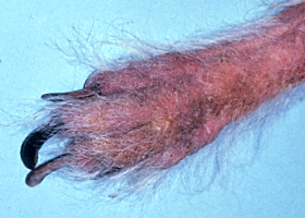
hemorrhagic bullae
Blood-filled elevation of epidermis, >1cm
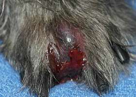
Interdigital hemorrhagic bulla in a dog with deep pyoderma and furunculosis
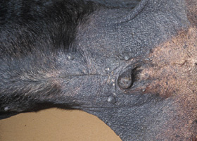
comedo
dilated hair follicle filled with keratin, sebum
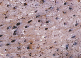
Comedones on the ventral abdomen of a dog with hypercortisolism
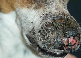
crust
Dried exudate and keratinous debris on skin surface
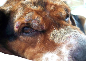
Multifocal crusts due to pemphigus foliaceus
epidermal collarettes
Circular scale or crust with erythema, associated with folliculitis or ruptured pustules or vesicles
Epidermal collarettes in a dog with Staphylococcus superficial bacterial folliculitis
erosion
Defect in epidermis that does not penetrate basement membrane. Histopathology may be needed to differentiate from ulcer.
Erosions in a dog with vasculitis
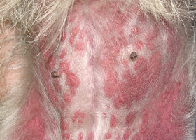
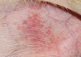
erythema
Red appearance of skin due to inflammation, capillary congestion
Erythema in a dog with cutaneous drug eruption
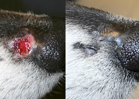
eschar
Thick crust often related to necrosis, trauma, or thermal/chemical burn
Eschar from physical trauma
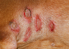
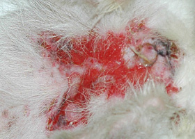
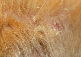
excoriation
Erosions and/or ulcerations due to self-trauma
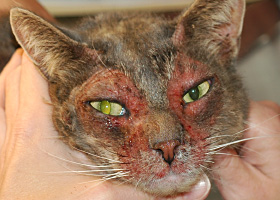
Excoriations in a cat with atopic dermatitis
fissure
Excessive stratum corneum, confirmed via histopathology. This term is often used to describe the nasal planum and footpads.
Fissures of the footpads in a dog with superficial necrolytic dermatitis
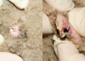
fistula
Ulcer on skin surface that originates from and is contiguous with tracts extending into deeper, typically subcutaneous tissues
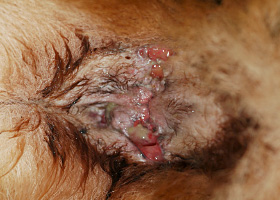
Perianal fistulas in a dog
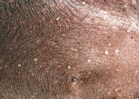
follicular casts
Accumulation of scale adherent to hair shaft
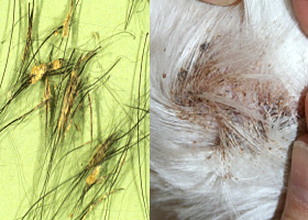
Follicular casts surrounding hairs from a dog with hypothyroidism
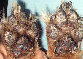
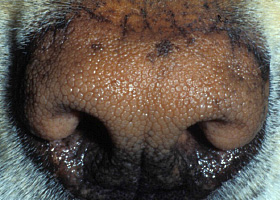
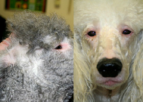
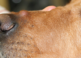
hyperkeratosis
Excessive stratum corneum, confirmed via histopathology. This term is often used to describe the nasal planum and footpads.
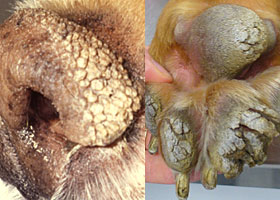
Idiopathic hyperkeratosis of the nasal planum (left) and footpads (right)
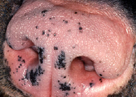
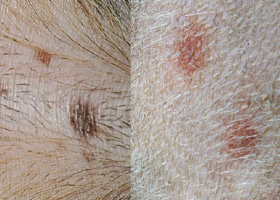
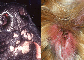
hyperpigmentation
Increased melanin in skin, often secondary to inflammation
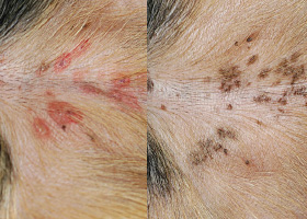
Inflammatory lesions (left) resulting in post-inflammatory hyperpigmentation (right)
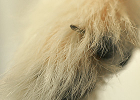
hypotrichosis
Lack of hair due to genetic factors or defects in embryogenesis.
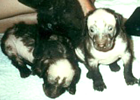
Congenital hypotrichosis in chocolate Labrador puppies.
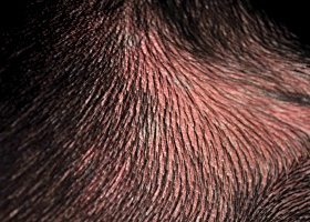
lichenification
Thickening of the epidermis, often due to chronic inflammation resulting in exaggerated texture
Lichenification of skin in a dog with chronic atopic dermatitis and Malassezia dermatitis
macule
Flat lesion associated with color change <1cm
Pigmented macule (left) Erythematous macule (right)
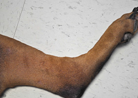
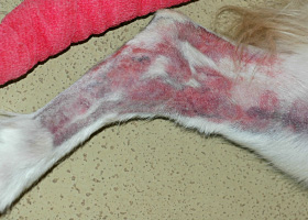
melanosis
Increased melanin in skin, may be secondary to inflammation.
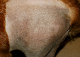
Post inflammatory hyperpigmentation of this dog’s thigh
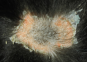
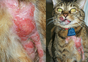
miliary
Multifocal, papular, crusting dermatitis; a descriptive term, not a diagnosis
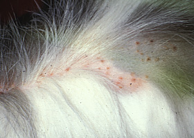
Miliary dermatitis in a flea allergic cat
morbiliform
A erythematous, macular, papular rash; the erythematous macules are typically 2-10 mm in diameter with coalescence to form larger lesions in some areas
Morbiliform eruptions in a dog with a cutaneous drug reaction
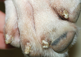
onychodystrophy
Abnormal nail morphology due to nail bed infection, inflammation, or trauma; may include: Onychogryphosis, Onychomadesis, Onychorrhexis, Onychoschizia
Onychodystrophy in dog with chronic allergies
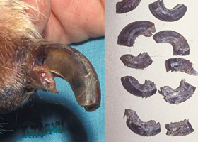
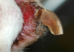
onychogryphosis
Abnormal claw curvature; secondary to nail bed inflammation or trauma
Onychogryphosis in a dog with symmetric lupoid onychodystrophy
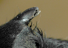
onychomadesis
Claw sloughing due to nail bed inflammation or trauma
Onychomadesis in a dog with symmetric lupoid onychodystrophy
onychorrhexis
Claw fragmentation due to nail bed inflammation or trauma
Onychorrhexis in a dog with symmetric lupoid onychodystrophy
onychoschizia
Claw splitting due to nail bed inflammation or trauma
Onychoschizia in a dog with symmetric lupoid onychodystrophy
patch
Flat lesion associated with color change >1cm
Hypopigmented patch (left), erythematous patch (right)
petechiae
Small erythematous or violaceous lesions due to dermal bleeding
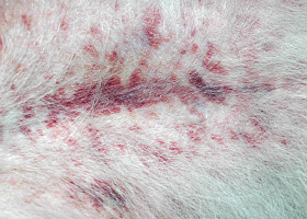
Petechiae in a dog with cutaneous vasculitis
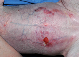
phlebectasia
Venous dilation; most commonly associated with hypercortisolism
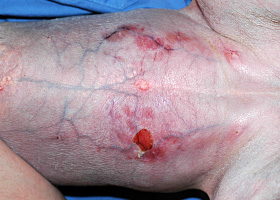
Phlebectasia and cutaneous atrophy due to hypercortisolism in a dog
plaques
Flat-topped elevation >1cm formed of coalescing papules or dermal infiltration
Plaques in a cat with cutaneous lymphoma
pustule
Raised epidermal infiltration of pus
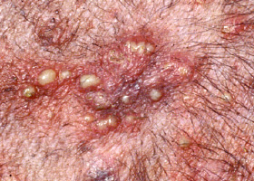
Pustules on the abdomen of a dog with superficial staphylococcal pyoderma.
scale
Accumulation of loose fragments of stratum corneum
Loose, large scales due to ichthyosis in a Golden Retriever
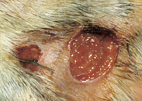
scar
Fibrous tissue replacing damaged cutaneous and/or subcutaneous tissues
Scarring (right) following the healing of an ulcer (left) in a dog with sterile nodular dermatitis
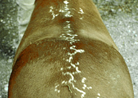
serpiginous
Undulating, serpentine (snake-like) arrangement of lesions
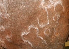
Serpiginous urticarial lesions on a horse
telangiectasia
Permanent enlargement of vessels resulting in a red or violet lesion (rare)
Telangiectasia in a dog with angiomatosis
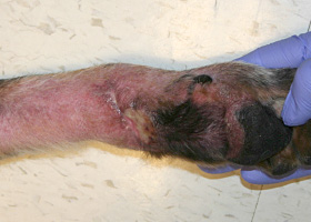
ulcer
A defect in epidermis that penetrates the basement membrane. Histopathology may be needed to differentiate from an erosion.
Ulcerations of the skin of a dog with vasculitis.
urticaria
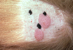
Wheals (steep-walled, circumscribed elevation in the skin due to edema ) due to hypersensitivity reaction
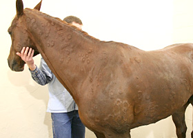
Urticaria in a horse
vesicle
Fluid-filled elevation of epidermis, <1cm
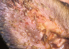
Vesicles and bullae on ear pinna due to bullous pemphigoid
wheal
Steep-walled, circumscribed elevation in the skin due to edema
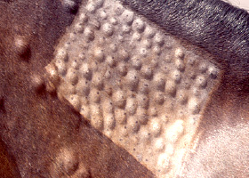
Wheals associated with intradermal allergy testing in a horse