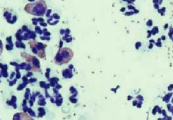
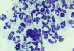
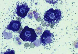
皮膚細胞診
いつ行うか?
- 細菌感染または酵母菌感染が疑われる場合(炎症性脱毛、脂漏症、鱗屑、丘疹、膿疱、痂皮、びらん、潰瘍)
- 結節/腫瘍のある患者-すべての結節/腫瘍に細胞診を実施する。
- 天疱瘡様疾患の疑いのある患者(びらん、膿疱、痂皮)
- 外耳炎のあるすべての患者
何を見つけるか?
- 球菌(ブドウ球菌属である可能性がもっとも高い)
- 桿菌 →培養および感受性試験が推奨される。
- 炎症性細胞の細胞内にみられる細菌→抗菌薬の全身投与が必要となる可能性のある、臨床的に重大な感染症である。
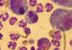
- 好酸球→外寄生生物またはアレルギーを示している可能性がある。
- マクロファージ→慢性の無菌性および感染性プロセスでみられる。
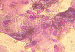
- マラセチア属真菌 → 油浸視野(倍率1,000倍)あたり1個以上のマラセチア属真菌は臨床的に重大と考えられる(正常な数は気候により変動する)マラセチア属過敏症の場合、はるかに少ない数の細菌(例、2または3 HPFあたり1個)でも臨床徴候を引き起こす可能性がある。
外用療法または全身療法を検討する必要がある。 - 腫瘍性細胞

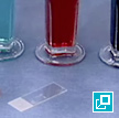
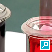
何が必要か?
- スライド、DiffQuick®または同等の染色液、ミネラルオイル、粘着テープ、顕微鏡、針、シリンジ
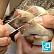
どのように行うか?
- 滲出液により湿り気のある感染皮膚表面か、脂を含んだ感染皮膚表面にスライドをこすりつけるか押しつける。
- 綿棒を皮膚表面で転がすか、耳の中に差し入れた後、スライド上で転がす。
- 膿疱のなかに針(25~27ゲージ)を皮膚に並行に差し入れ、膿疱のみに穴を開ける。深部細胞または血液の採取は必要ではない。膿疱頂部を持ち上げて外し、破裂した膿疱にスライドを押しつける。
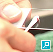
- 粘着テープの粘着面を用いて、乾燥し落屑した皮膚またはそのいずれか一方から細胞および表面組織を採取し、これをガラススライド上に置いて(粘着面を下向きに)、DiffQuick®の青い染色液を1滴垂らす。
- テープ自体がカバーガラスとなる。
スライドに両面粘着テープを貼り付け、粘着面で材料を採取する。これをDiffQuick®の青い染色液に浸漬し、乾燥させ、油浸下で調査する。
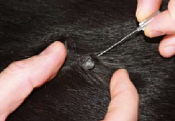
- 結節または膿瘍に針を挿入し、皮膚に接触させたまま数回入れ直す。針を引き抜く。この針に、内筒が引いてあるシリンジを取り付け、針の内容物をスライドに押し出して、空気乾燥させる。
- 空気乾燥したスライドを染色する(例、DiffQuick,sup>®)。
- スライドを顕微鏡下において、コンデンサを上げる。
ヒント
- 綿棒を生理食塩水で湿らせるか、スライドの端を慎重に皮膚にこすりつけ、次に材料をスライドにこすりつける。
- 透明な粘着テープ(粘着側を下向きに)を皮膚に押しつける。このテープをスライドのように染色し、空気乾燥させてからスライドに押しつけるか、DiffQuick®の青い染色液をスライドに垂らし、粘着側を下にテープを染色液に押しつける。顕微鏡下で評価する。
abscess
A discrete swelling containing purulent material, typically in the subcutis
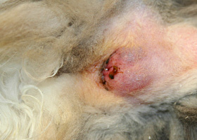
Perianal abscess in a dog
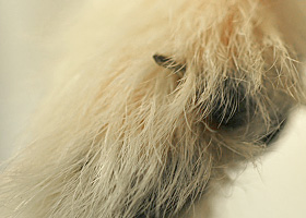
alopecia
Absence of hair from areas where it is normally present; may be due to folliculitis, abnormal follicle cycling, or self-trauma
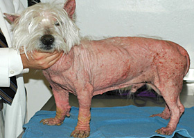
Extensive alopecia secondary to cutaneous epitheliotropic lymphoma
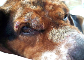
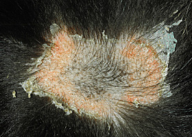
alopecia (“moth-eaten”)
well-circumscribed, circular, patchy to coalescing alopecia, often associated with folliculitis
“Moth-eaten” alopecia secondary to superficial bacterial folliculitis
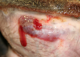
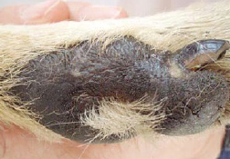
hemorrhagic bullae
Blood-filled elevation of epidermis, >1cm
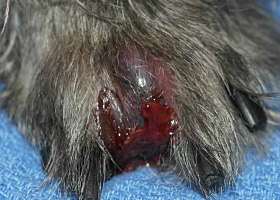
Interdigital hemorrhagic bulla in a dog with deep pyoderma and furunculosis
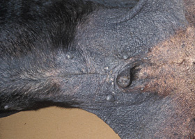
comedo
dilated hair follicle filled with keratin, sebum
Comedones on the ventral abdomen of a dog with hypercortisolism
crust
Dried exudate and keratinous debris on skin surface
Multifocal crusts due to pemphigus foliaceus
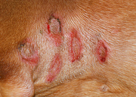
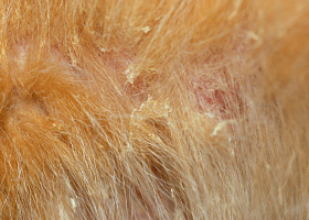
epidermal collarettes
Circular scale or crust with erythema, associated with folliculitis or ruptured pustules or vesicles
Epidermal collarettes in a dog with Staphylococcus superficial bacterial folliculitis
erosion
Defect in epidermis that does not penetrate basement membrane. Histopathology may be needed to differentiate from ulcer.
Erosions in a dog with vasculitis
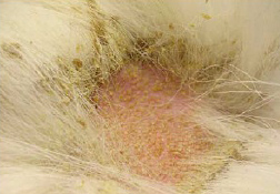
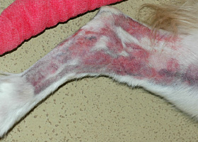
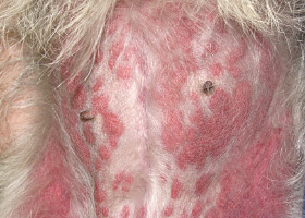
erythema
Red appearance of skin due to inflammation, capillary congestion
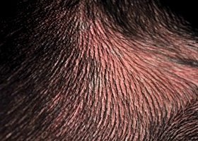
Erythema in a dog with cutaneous drug eruption
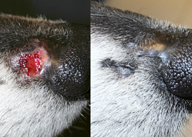
eschar
Thick crust often related to necrosis, trauma, or thermal/chemical burn
Eschar from physical trauma
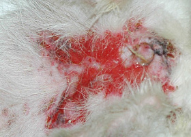
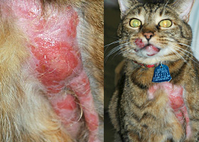
excoriation
Erosions and/or ulcerations due to self-trauma
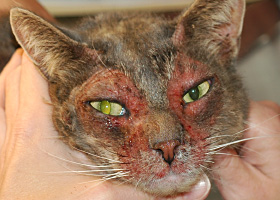
Excoriations in a cat with atopic dermatitis
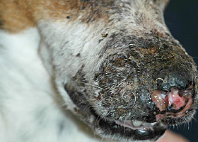
fissure
Excessive stratum corneum, confirmed via histopathology. This term is often used to describe the nasal planum and footpads.
Fissures of the footpads in a dog with superficial necrolytic dermatitis
fistula
Ulcer on skin surface that originates from and is contiguous with tracts extending into deeper, typically subcutaneous tissues
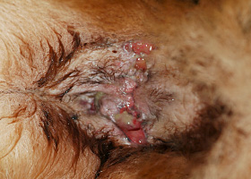
Perianal fistulas in a dog
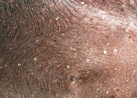
follicular casts
Accumulation of scale adherent to hair shaft
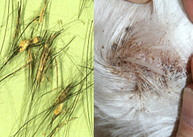
Follicular casts surrounding hairs from a dog with hypothyroidism
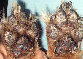
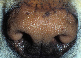
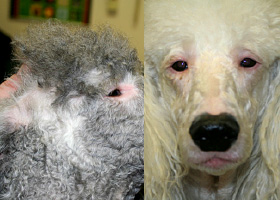
hyperkeratosis
Excessive stratum corneum, confirmed via histopathology. This term is often used to describe the nasal planum and footpads.
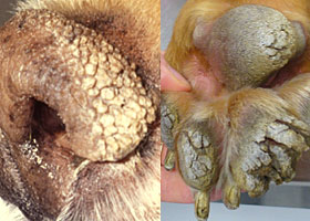
Idiopathic hyperkeratosis of the nasal planum (left) and footpads (right)
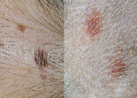
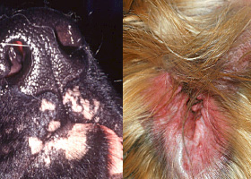
hyperpigmentation
Increased melanin in skin, often secondary to inflammation
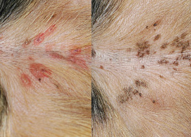
Inflammatory lesions (left) resulting in post-inflammatory hyperpigmentation (right)
hypotrichosis
Lack of hair due to genetic factors or defects in embryogenesis.
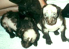
Congenital hypotrichosis in chocolate Labrador puppies.
lichenification
Thickening of the epidermis, often due to chronic inflammation resulting in exaggerated texture
Lichenification of skin in a dog with chronic atopic dermatitis and Malassezia dermatitis
macule
Flat lesion associated with color change <1cm
Pigmented macule (left) Erythematous macule (right)
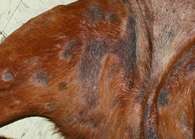
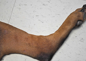
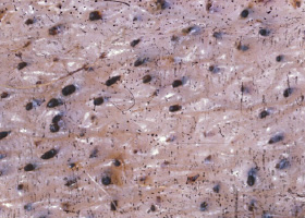
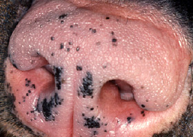
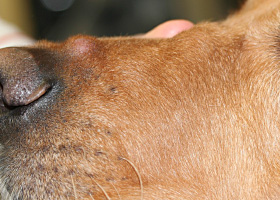
melanosis
Increased melanin in skin, may be secondary to inflammation.
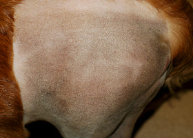
Post inflammatory hyperpigmentation of this dog’s thigh
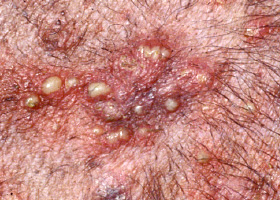
miliary
Multifocal, papular, crusting dermatitis; a descriptive term, not a diagnosis
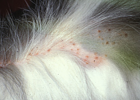
Miliary dermatitis in a flea allergic cat
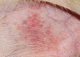
morbiliform
A erythematous, macular, papular rash; the erythematous macules are typically 2-10 mm in diameter with coalescence to form larger lesions in some areas
Morbiliform eruptions in a dog with a cutaneous drug reaction
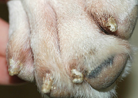
onychodystrophy
Abnormal nail morphology due to nail bed infection, inflammation, or trauma; may include: Onychogryphosis, Onychomadesis, Onychorrhexis, Onychoschizia
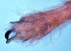
Onychodystrophy in dog with chronic allergies
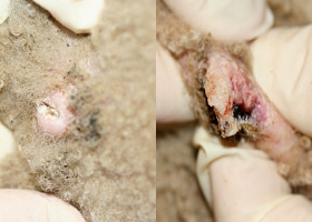
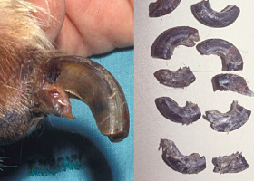
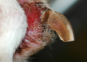
onychogryphosis
Abnormal claw curvature; secondary to nail bed inflammation or trauma
Onychogryphosis in a dog with symmetric lupoid onychodystrophy
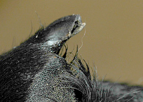
onychomadesis
Claw sloughing due to nail bed inflammation or trauma
Onychomadesis in a dog with symmetric lupoid onychodystrophy
onychorrhexis
Claw fragmentation due to nail bed inflammation or trauma
Onychorrhexis in a dog with symmetric lupoid onychodystrophy
onychoschizia
Claw splitting due to nail bed inflammation or trauma
Onychoschizia in a dog with symmetric lupoid onychodystrophy
patch
Flat lesion associated with color change >1cm
Hypopigmented patch (left), erythematous patch (right)
petechiae
Small erythematous or violaceous lesions due to dermal bleeding
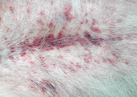
Petechiae in a dog with cutaneous vasculitis
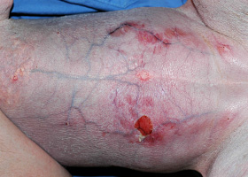
phlebectasia
Venous dilation; most commonly associated with hypercortisolism
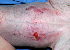
Phlebectasia and cutaneous atrophy due to hypercortisolism in a dog
plaques
Flat-topped elevation >1cm formed of coalescing papules or dermal infiltration
Plaques in a cat with cutaneous lymphoma
pustule
Raised epidermal infiltration of pus
Pustules on the abdomen of a dog with superficial staphylococcal pyoderma.
scale
Accumulation of loose fragments of stratum corneum
Loose, large scales due to ichthyosis in a Golden Retriever
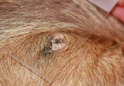
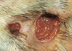
scar
Fibrous tissue replacing damaged cutaneous and/or subcutaneous tissues
Scarring (right) following the healing of an ulcer (left) in a dog with sterile nodular dermatitis
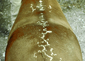
serpiginous
Undulating, serpentine (snake-like) arrangement of lesions
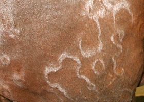
Serpiginous urticarial lesions on a horse
telangiectasia
Permanent enlargement of vessels resulting in a red or violet lesion (rare)
Telangiectasia in a dog with angiomatosis
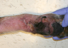
ulcer
A defect in epidermis that penetrates the basement membrane. Histopathology may be needed to differentiate from an erosion.
Ulcerations of the skin of a dog with vasculitis.
urticaria
Wheals (steep-walled, circumscribed elevation in the skin due to edema ) due to hypersensitivity reaction
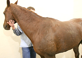
Urticaria in a horse
vesicle
Fluid-filled elevation of epidermis, <1cm
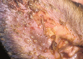
Vesicles and bullae on ear pinna due to bullous pemphigoid
wheal
Steep-walled, circumscribed elevation in the skin due to edema
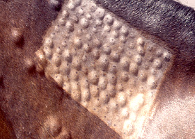
Wheals associated with intradermal allergy testing in a horse